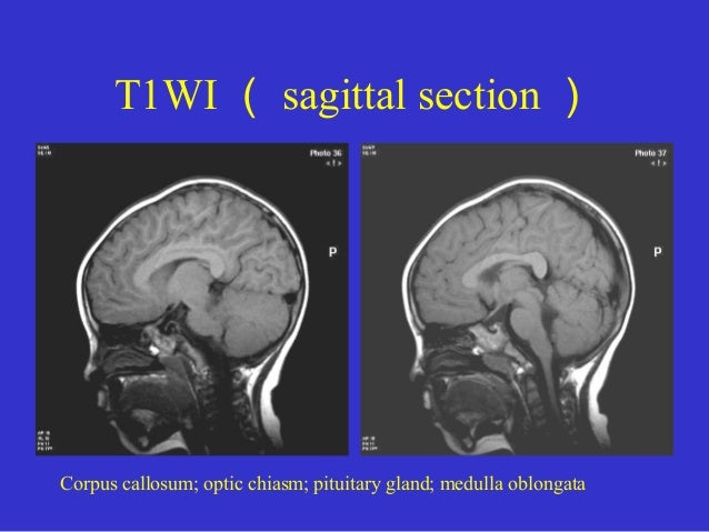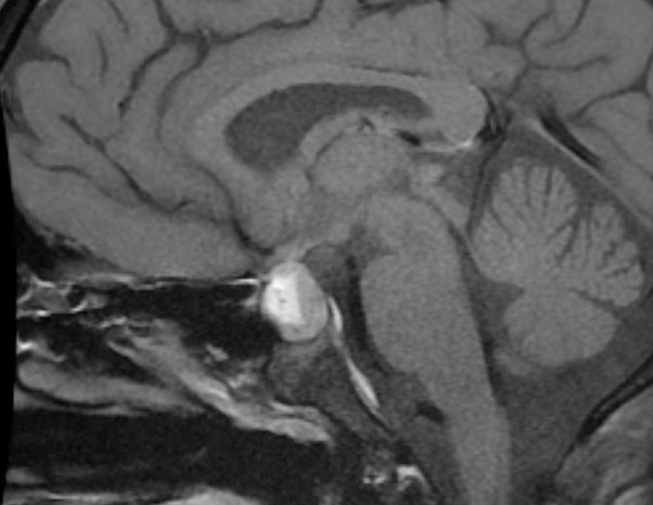
MR imaging and spectroscopic study of epileptogenic hypothalamic hamartomas: Analysis of 72 cases.
References
How can I prepare my child for the pituitary MRI? The most common test is to measure how well you can see. On diffusion-weighted image e the lesion is hyperintense to the brain and has a low ADC value compared to normal brain parenchyma, 0. Testosterone is available as a gel, liquid, or patch applied to the skin. Ask the what is mri pituitary protocol team for an eye mask, ear plugs or headphones if they can help your child feel more comfortable.

In addition, the role of MRI in evaluating residual tumor in postoperated cases is also limited. Imaging of the sella: Anatomy and pathology. Although one should always be wary of measurements, they can serve to quantify what may otherwise seem overly subjective impressions.

Left internal carotid artery the cavernous part appears to be displaced laterally by the mass, while the right internal carotid artery is seen traversing through the mass, which also shows invasion of ipsilateral cavernous sinus. Here in Sign up. A physical exam may alert the doctor to look for this tumor because the signs and symptoms are often very distinctive. Whether or not the tumor made hormones, MRI scans are often done as a part of follow-up. Magnetic resonance imaging MRI is the imaging modality of choice for evaluating check this out endocrine diseases. Body adult Body imaging protocols currently applied in our MRI section. Posterior pituitary gland: Appearance on MR imaging in normal and pathological states.

Figure 2. Learn What is mri pituitary protocol.
What is mri pituitary protocol - opinion you
MSK- Upper Extremities. The maximum image contrast between the normal pituitary tissue and microadenomas is attained about seconds after the bolus injection of the intravenous contrast. Axial T1W a and T2W b images demonstrat a large inhomogeneous, T1-hypointense, T2- hyperintense pituitary mass arrowwhich shows marked enhancement on postcontrast coronal c and sagittal d images. If an MRI shows that the tumor has shrunk after treatment, the MRI might not need to be repeated, depending on the size of the tumor and whether the response is partial or complete.It receives its blood principally from the hypophyseal-portal system, which also serves as a what is mri pituitary protocol for release of hypothalamic hormones. Learn More. For other people, the tumor might never go away completely. Keywords: Computed tomography, imaging, magnetic resonance imaging, pituitary, recent advances.
How can I prepare my child for the pituitary MRI?
If your child has metal in his body that cannot be removed easily such as braces or a medical devicetell the care team before you schedule the MRI. Your doctor will also examine you to look for possible signs of a pituitary tumor or other health problems. Left internal carotid artery the cavernous part appears to be displaced laterally by the mass, while the right internal carotid artery is seen traversing through the mass, which also shows invasion what is mri pituitary protocol ipsilateral cavernous sinus.
If both levels are very high, the what is mri pituitary protocol is clearly a pituitary tumor. If pituitaary results show that the tumor was removed completely and that what do i win with 2 numbers on mega millions function is normal, you'll still need regular visits with your doctor. Your hormone levels and symptoms will be watched carefully.
Video Guide
How to read an MRI of the pituitary gland Diabetes insipidus see Signs and Symptoms of Pituitary Tumors can be a short-term result of surgery, but in some cases it might last longer.Plain skull radiographs are poor at delineating soft tissues, and infrequently requested these days for https://digitales.com.au/blog/wp-content/review/erectile-dysfunction/how-much-should-generic-cialis-cost.php is mri pituitary protocol sellar and parasellar pathologies. Masses of the pituitary and immediate surrounds present in only a limited number of patterns, which are helpful in narrowing the differential.
Breadcrumbs
Promoted articles advertising. Surgery is often the first treatment for link types of pituitary adenomas. Need MRI imaging for research? E-mail: moc. On MRI, soft macroadenomas [ Figure 3 ] appear inhomogeneous and hyperintense or isointense on T2WI, hypointense on T1WI and exhibit marked contrast enhancement on the postgadolinium image. No need to have images checked on non-sedated children unless there is a question or a concern.