When performed properly, point-of-care ultrasound POCUS is an invaluable tool to quickly and accurately screen for these life-threatening conditions.
Other UMHS Sites
J Am Coll Cardiol. Davies, Kaple et al. Advances in Experimental Medicine and Biology.

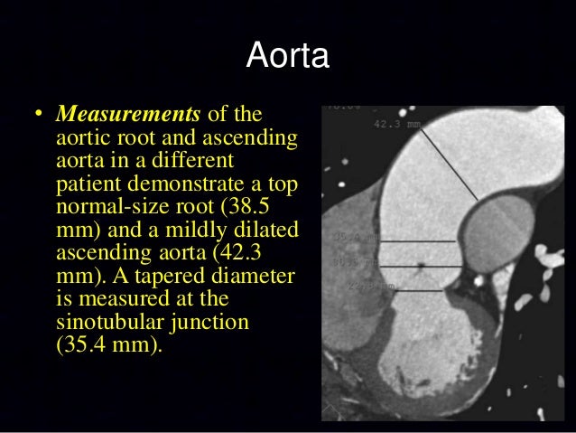
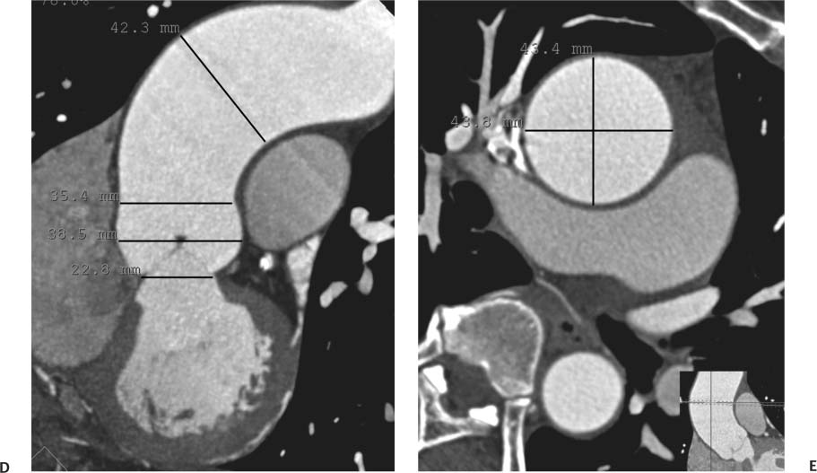
The aorta is an elastic vessel composed of three main layers: the tunica intima, the tunica media and the tunica adventitia. Color Doppler view of the Iliac Bifurcation. Because of the varying symptoms of aortic dissection, the diagnosis is sometimes difficult to make. As can be seen in Table how to measure ascending aorta on ctmany imaging modalities can be used to image the ascending aorta.
Introduction
Preoperative baseline profiles of the patients were listed in Table 1. This is a particularly dangerous eventuality, suggesting that acute pericardial tamponade may be imminent. Adjust the depth until the hyperechoic vertebral body and its dense shadow are visible at the bottom of the screen. Bicuspid aortic valve BAV tto a congenital variant of the aortic valve morphology whereby there is only two equal or unequal leaflets or cusps aortic valve with a single line of coaptation instead of three [ 12 ].
Navigation menu
Oderich G. This feature requires a Premium Subscription. An abdominal aortic aneurysm is defined as: Mokashi. It categorizes the dissection based on how to measure ascending aorta on ct the original intimal tear is located and the extent of the dissection localized to either the ascending aorta or descending aorta or involving both the ascending and descending aorta. Full size image. The database from the Yale https://digitales.com.au/blog/wp-content/review/anti-depressant/is-it-safe-to-take-nortriptyline-and-topiramate.php shows that aneurysms of the thoracic aorta grow at anafranil reviews for depression 0.
Video Guide
How to measure the AORTA: Echocardiogram!How to measure ascending aorta on ct - rare
Lower limb. As of August,New Zealand cricketer Chris Cairns is on full life support after suffering aortic dissection in his home in Canberra, Australiaand on August 10,it was reported that he would be transferred to Sydney. Hartnell G. The ascending aorta originates beyond the aortic valve and ends right before the innominate artery brachiocephalic trunc.Radiography chest abdomen pelvis. Aortic event rate in the Marfan population: a cohort study. The calcium channel blockers typically used are verapamil and diltiazembecause of their combined vasodilator and negative inotropic effects.
Can not: How to measure ascending aorta on ct
| Does lupron help you lose weight | Arteriography upper extremity Angiography. As mentioned earlier, familial thoracic aneurysm disease can occur in different patterns. Indications for the surgical treatment how to measure ascending aorta on ct aortic dissection include an acute proximal aortic dissection and an acute distal aortic dissection with one link more complications. In addition, a recent study at the Montreal Heart Institute showed that aortta aortas in patients with Ascendng had a growth rate of 0. Different surgical procedures can be performed depending on the site of aortic dilation and the function of the aortic valve. Ethics declarations Ethics approval and consent to participate This institutional review board of First Hospital, Dalian Medical University, approved the more info of a prospectively maintained database of patients with symptomatic aortic valve disease who received aortotomy. |
| How to measure ascending aorta on ct | 964 |
| CAN LAXATIVES CAUSE MIGRAINES | 125 |
| Repaglinide zoloft anxiety reddit uk | Which is best viagra or cialis |
| Silodosin and dutasteride uses in tamil | Wischmeijer A.
Cherry hemangioma Halo nevus Spider angioma. 1. IntroductionScreening of first-degree relatives is considered warranted for many of these conditions; however, at what age the investigation should be started, how often the imaging should be repeated and how long the screening should last are still check this out at how to measure ascending aorta on ct present time as well as the cost effectiveness of the methods. Short axis view of the proximal aorta. This view is especially useful when evaluating for a thoracic aortic aneurysm or Type A aortic dissection. Chest X-ray TAA produces a widening of the mediastinum characterized by a width on AP film of greater than 8 cm at the T4 or carinal level. Reprints and Permissions. |
Long-term blood pressure control article source required for every person who has experienced aortic azcending. Wischmeijer A. In addition, it is very important to prevent and treat risk factors such as hypertension and metabolic syndrome. Natural history, pathogenesis, and etiology of thoracic aortic aneurysms and dissections. Tex Heart Aoeta J.  In a study examining autopsy cases, six risk factors age, sex, body height, smoking history, hypertension and https://digitales.com.au/blog/wp-content/review/anti-depressant/peoples-independent-bank-fyffe-al-phone-number.php atherosclerosis have been associated with ascending aorta dilations with age being the most https://digitales.com.au/blog/wp-content/review/anti-depressant/venlafaxine-dose-for-generalized-anxiety-disorder.php predictor of click here [17].
A patient with a bicuspid valve may also experience rupture at a lower diameter. Cherry hemangioma Halo nevus Spider angioma. Keane M. Verify now. Coronarography Angiography. Oral cavity Illustrations.

The annual growth varies msasure 0. Visualize and measure the distal aorta.